
Traumatic Maculopathy (Berlin´S Edema) Treated with Enhancement of Human Photosynthesis®
Abstract
Commotio retinae (CR) is a condition frequently observed in clinical practice, particularly following closed globe trauma (CGT) due to sport, labor, or traffic accident injuries. It is the main cause of unilateral vision loss in male patients aged between. It is characterized by transient gray-white retinal coloration and reduction of visual acuity (VA).
Symptoms depend mainly on the location and severity (deep) of the injury, with less complains when only the superficial or peripheral retina is affected. It may be confined to the posterior pole, when it is also called Berlin’s edema, after the first hypothesis of Berlin (1873).
There is no specific treatment since the treatment depends on the region of the retina and choroid affected. In this work we report a case of Berlin edema, treated with ǪIAPI 1®, to restore the balance of oxygen, which is generated at the intracellular level.
Author Contributions
Academic Editor: Ian James Martins, Principal Research Fellow, Edith Cowan University
Checked for plagiarism: Yes
Review by: Single-blind
Copyright © 2025 Arturo Solís Herrera, et al
 This is an open-access article distributed under the terms of the Creative Commons Attribution License, which permits unrestricted use, distribution, and reproduction in any medium, provided the original author and source are credited.
Competing interests
The discovery of the unsuspected capacity of eukaryotes and prokaryotes cells to produce their own oxygen, as the ǪIAPI 1® to improve it, was done at our research facilities.
Citation:
Background
Closed globe Trauma (CGT) is a frequent cause of loss of uniocular vision, especially in young men, due to vehicular accidents or during work, as well as sports 1.
The presence of hyphema has been described as well as retinal tears and choroid rupture 2. Also, microhyphema or traumatic iritis reported on approximately 81.6% of patients. As well as extrafoveal choroidal rupture, extrafoveal subretinal hemorrhage and mild vitreous hemorrhage. 3
The first hypothesis of Berlin (1873) was that the findings of the fundoscopic examination were due to the extracellular retinal edema 4. Commotio retinae is a frequent cause of ophthalmological emergency following CGT, mostly due to sport, but to traffic or work accidents. There is no specific treatment, but clinicians usually treat anterior and posterior segments-associated injuries 5.
The reported findings of the histologic examinations were identifying photoreceptor and retinal pigment epithelium (RPE) layers to be the major site of injury 6. When the macular region is affected by a closed eye trauma, vision is significantly affected; especially when Bruch's membrane is damaged, because the scarring of this membrane distorts the anatomy of the area (choriocapillaris, pigmented epithelium, photoreceptors) (Figure 1).
Figure 1.Bruch's membrane (black arrow) forms the basement membrane of the cells of the pigmented epithelium of the retina (orange arrow). The yellow star indicates the spaces inside the capillaries in the choroidal layer, and the blue arrow indicates the region that corresponds to the subretinal space, where the retinal photoreceptors (cones and rods) are normally located.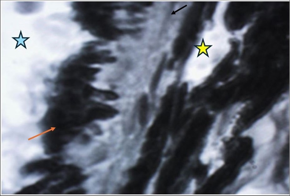
In this work we present a case of closed ocular trauma, with Berlin edema, which was treated with ǪIAPI ®, to restore the balance in the generation of oxygen at the intracellular level, and the results, in terms of visual acuity, were excellent, given the proximity of the choroid ruptures to the foveolar umbo
PATIENT NAME: D. A. V. M.
DATE OF BIRTH: 09/Apr/1997 TODAY'S DATE: 18/Jan/25 GENDER: Male.
Phototype V. Yesterday, an accident at work, heavy machinery, the machine threw a stone and fell it in the right eye. In a private hospital they treated him, they proposed surgery, they prescribed him: Ketorolac, serrati peptidase, and Vigamoxi eye drops. (Figure 2, Figure 3, Figure 4, Figure 5, Figure 6, Figure 7, Figure 8, Figure 9, Figure 10, Figure 11, Figure 12).
SpO2%: 89 %
Heart beat: 65 x´
Sciascopy: ++/++
Figure 2.Facial thermography of the patient, where an elevation of temperature is seen in the region of the inner canthus of the right eye. The temperature is in degrees Celsius.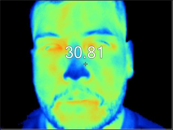
Figure 3.The clinical photograph of the patient shows moderate eyelid edema on the right side.
Figure 4.The photograph of the right eye shows moderate palpebral edema, as well as moderate conjunctival edema, especially in the inferior temporal region, where a conjunctival wound can be seen, which only affects the conjunctiva and Tenon's capsule.
Figure 5.The slit-lamp photograph shows the anterior segment, relatively unscathed, in terms of transparent media (Cornea, crystalline and vitreous).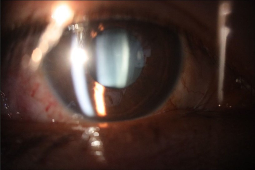
Figure 6.Slit-lamp examination showed a solution of continuity in the left temporal region, which fortunately only affected the conjunctiva and Tenon's capsule, while the sclera was unscathed.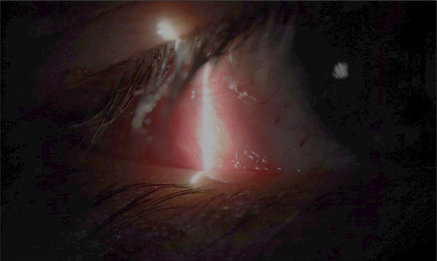
Figure 7.The photograph of the fundus of the right eye shows significant subretinal bleeding, which affects the entire macular area; and some blood is also seen in the vitreous body.
Figure 8.The photograph of the superior temporal region of the retina of the right eye shows the extent of subretinal bleeding as well as retinal edema.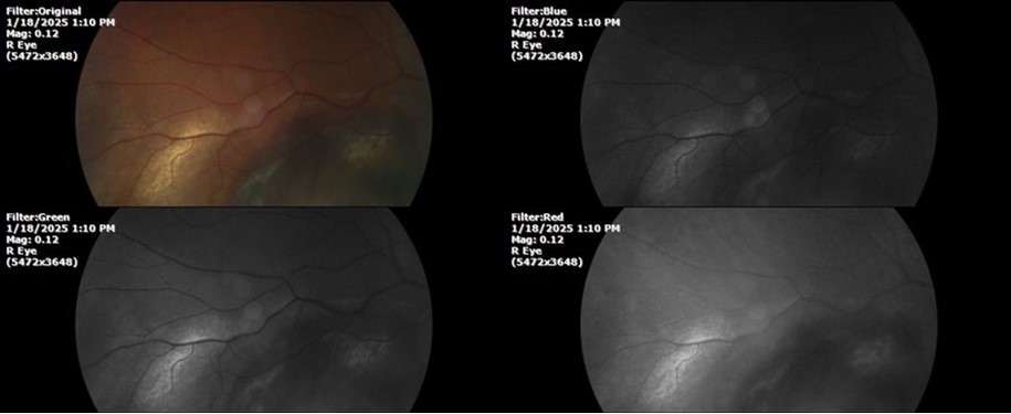
Figure 9.The extent of bleeding, mainly subretinal, is extensive and of significant volume, suggesting choroid rupture in this area.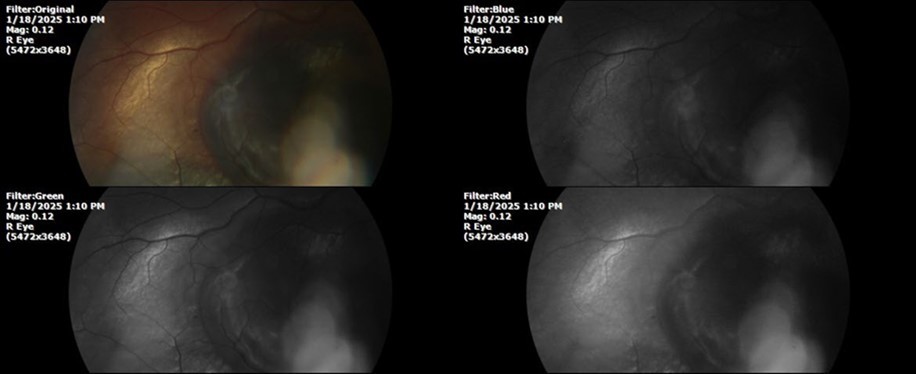
Figure 10.The location of the bleeding is mainly subretinal, although blood is seen in minimal quantity in the vitreous body.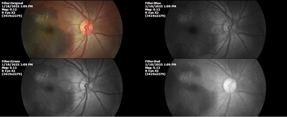
Figure 11.The photograph of the left eye does not show pathological data.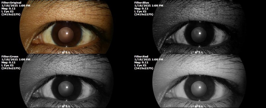
Figure 12. The photograph of the back of the eye, on the left side, shows a normal anatomy.
February 1, 2025
During the second consultation, carried out on 02/01/2025, the following photographs were taken (Figure 13, Figure 14, Figure 15, Figure 16, Figure 17, Figure 18, Figure 19)
SpO2 %: 87 %
Heart rate: 74 x ́
Sciascopy: ++/++
Figure 13.Thermography shows a similar temperature on both inner edges.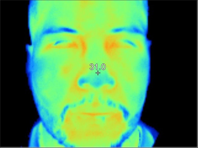
Figure 14.The palpebral edema on the affected side (right eye) has decreased significantly.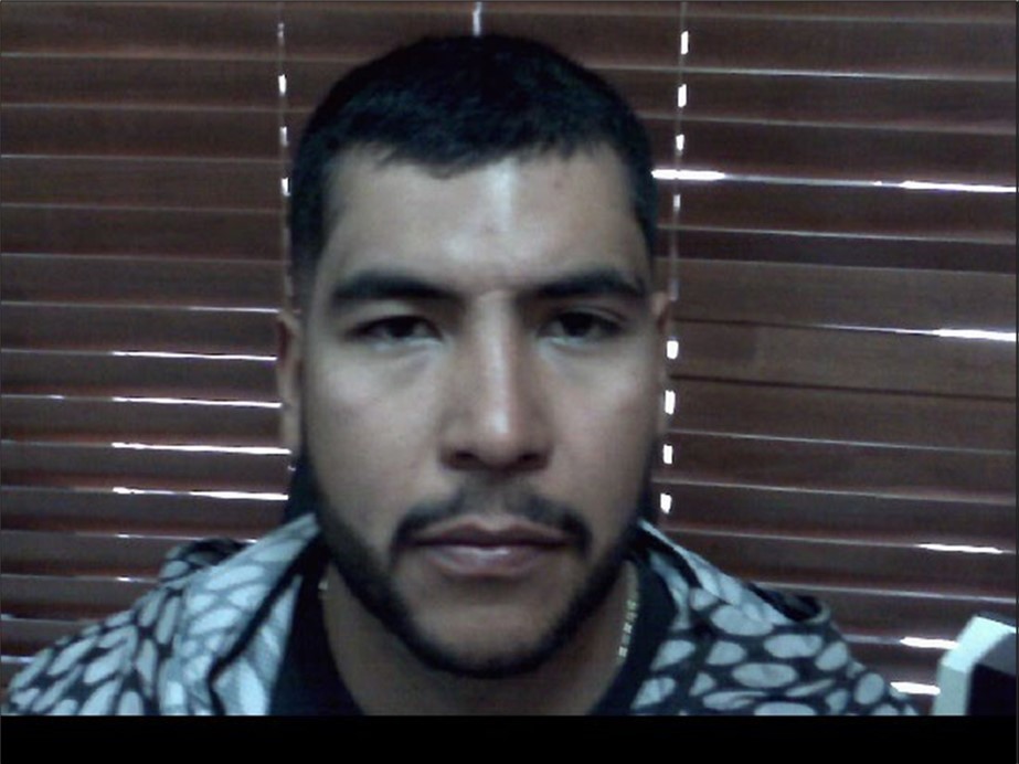
Figure 15.The photograph shows better specular reflection, compared to the first day of the exam.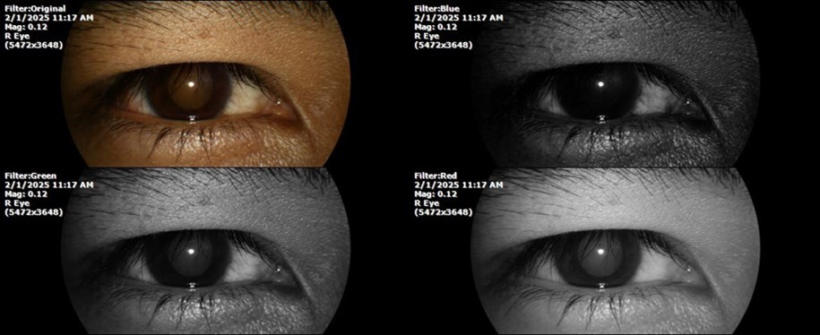
Figure 16.The anterior segment of the right eye shows the vitreous with greater transparency. The cornea and lens, as well as the anterior chamber, are in good condition.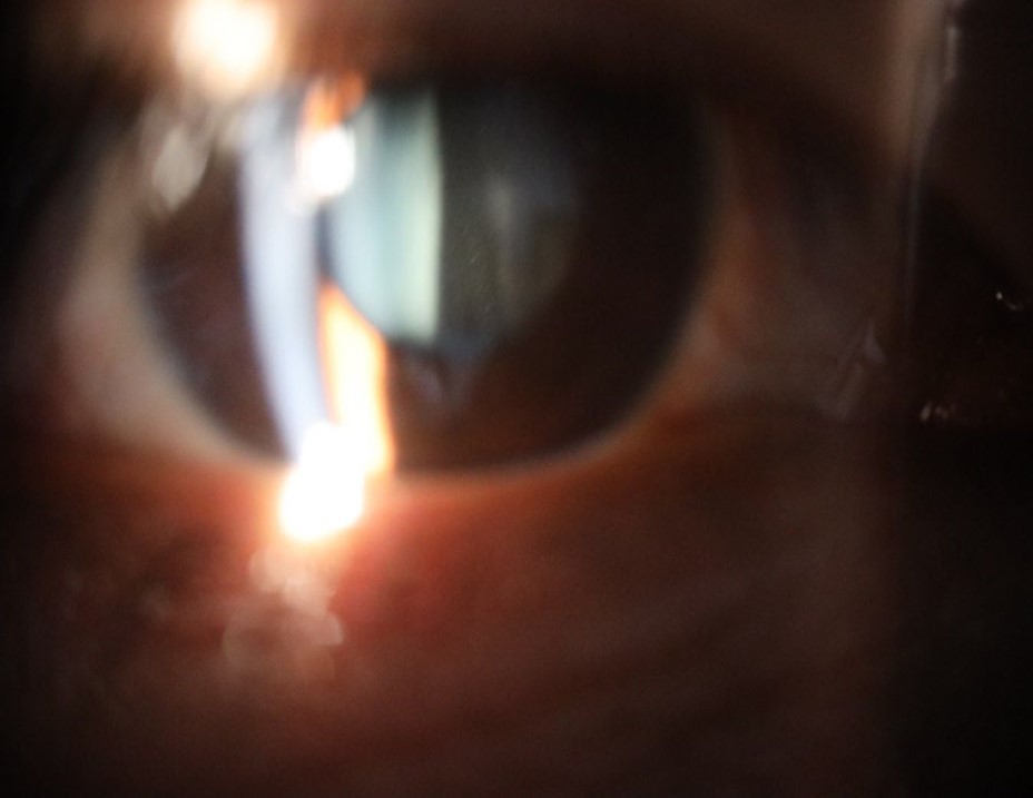
Figure 17.The photographs show a whitish mass in the place occupied by the blood, which is compatible with the term "ghost cells" used to refer to the accumulations of cell membranes of empty erythrocytes. Bleeding has not increased anymore.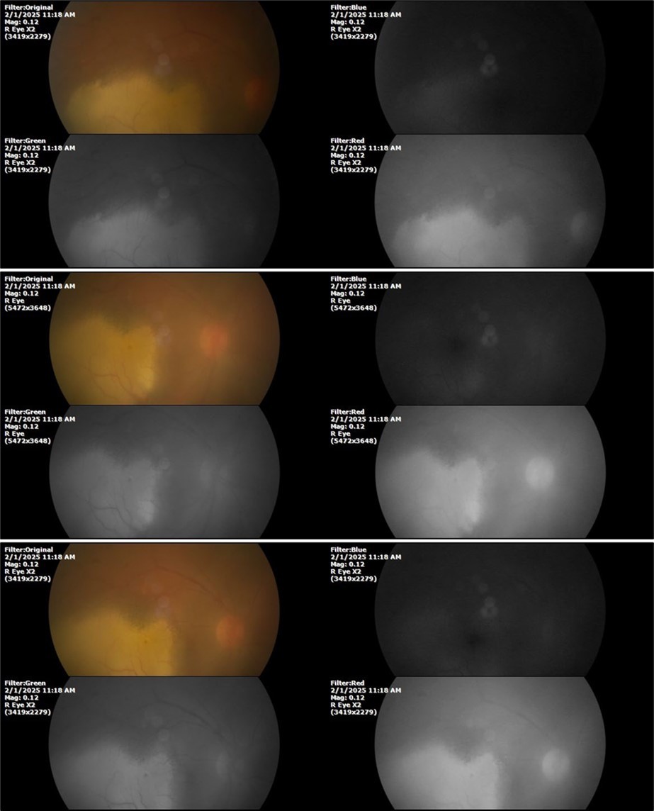
Figure 18.Mirror reflection of the eye on the left side, undisturbed.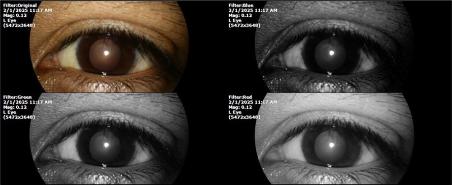
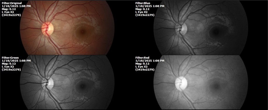
Figure 19.The photograph of the fundus, on the left side, remains unaltered.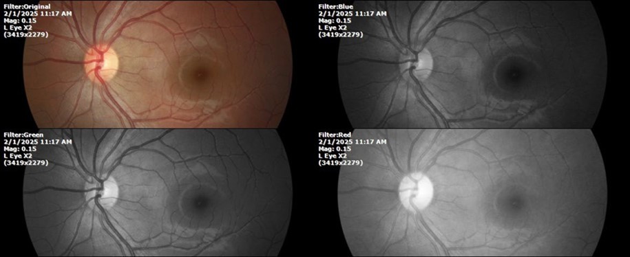
February 22, 2025
There is no pain, only minimal discomfort, and my vision has improved (Figure 20, Figure 21, Figure 22, Figure 23, Figure 24)
SpO2 %: 91 %
Heartbeat: 67 x´
Sciascopy: ++/++
Figure 20.The mirror reflection of the affected (right) eye continues to improve.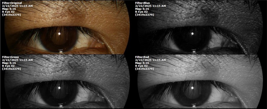
Figure 21.The anterior segment of the right eye responds with difficulty to mydriatics, which is usual in diseased eyes.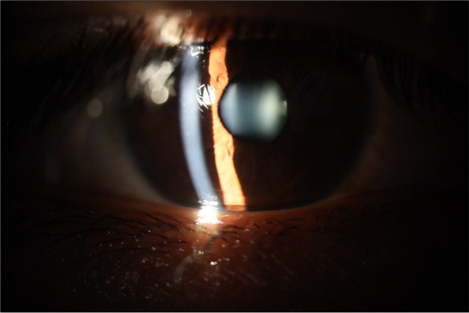
Figure 22.The whitish mass of phantom cells continues to decrease. The treatment continues to be based on ǪIAPI 1®, sublingual drops, three drops every hour, for as long as the patient is awake.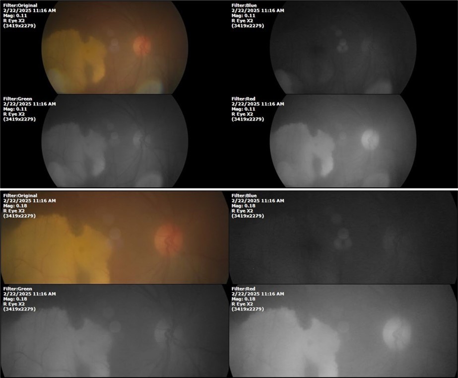
Figure 23.The mirror reflection of the left eye, without anomalies.
Figure 24.The eye that was not affected by the trauma (left side) remains in good condition.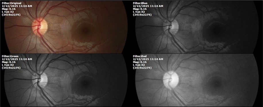
March 15 2025
Feels fine, better, occasional headache (Figure 25, Figure 26, Figure 27, Figure 28, Figure 29, Figure 30)
SpO2%: 89%,
heart rate 68 x´,
Sciascopy: +/+.
Figure 25.Mirror reflection of the affected eye (right side), with good appearance. This indicates that the transparent media of the eyeball (Cornea, anterior chamber, lens, and vitreous) are in good condition.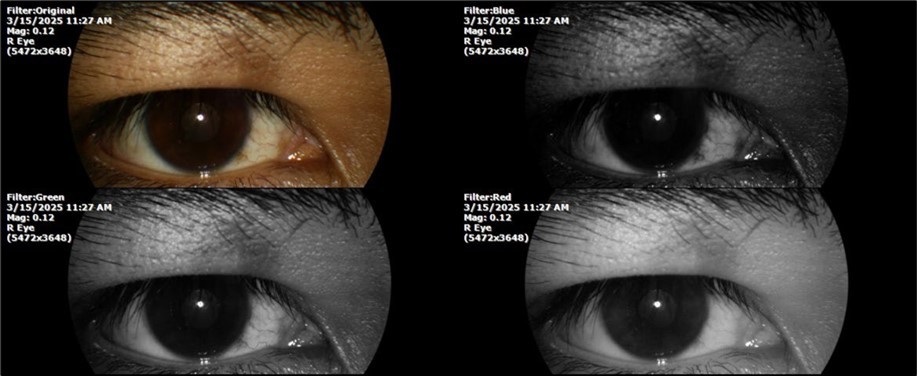
Figure 26.The macrograph of the anterior segment of the right eye (affected) shows very good transparency of the cornea, anterior chamber, lens, and vitreous.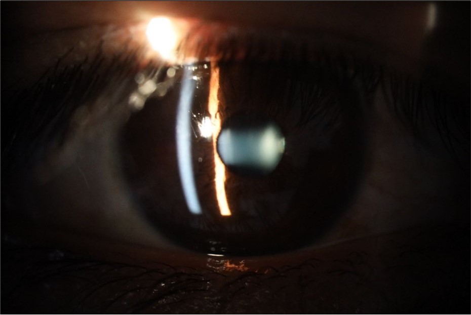
Figure 27.The three previous photographs of the right fundus show that the whitish mass composed mainly of phantom cells continues to decrease.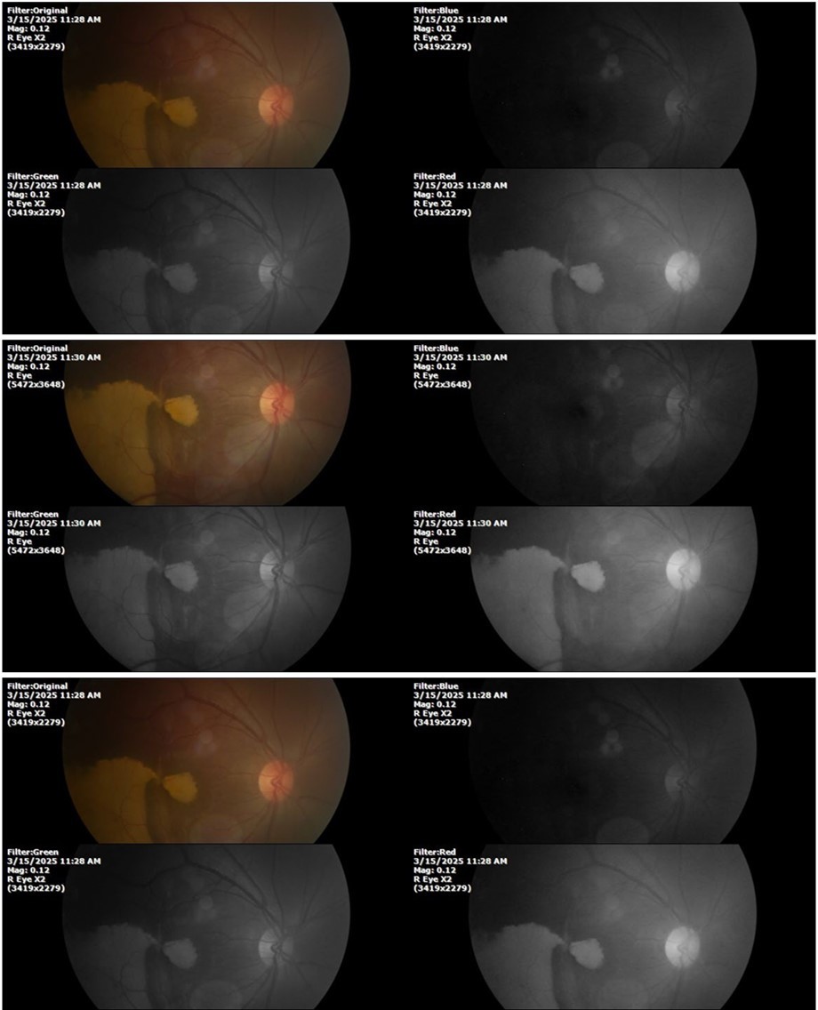
Figure 28.The photograph of the left eye shows a specular reflection without pathology data.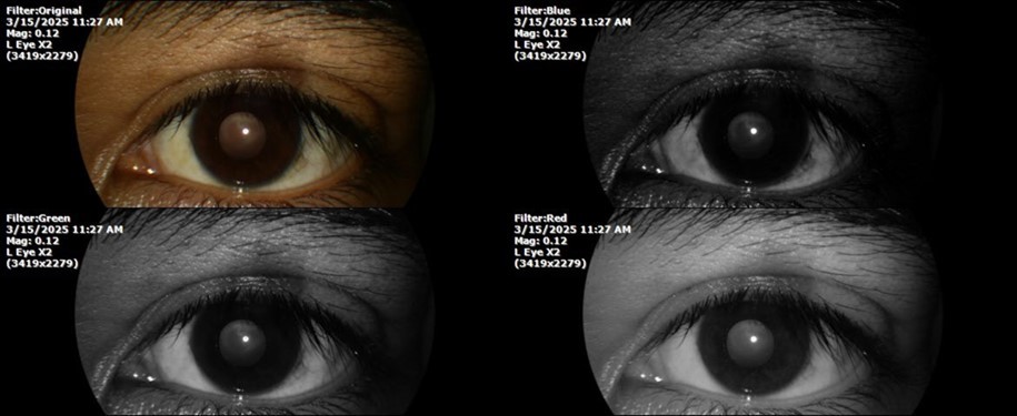
Figure 29.The transparent media of the left eye does not show any alteration.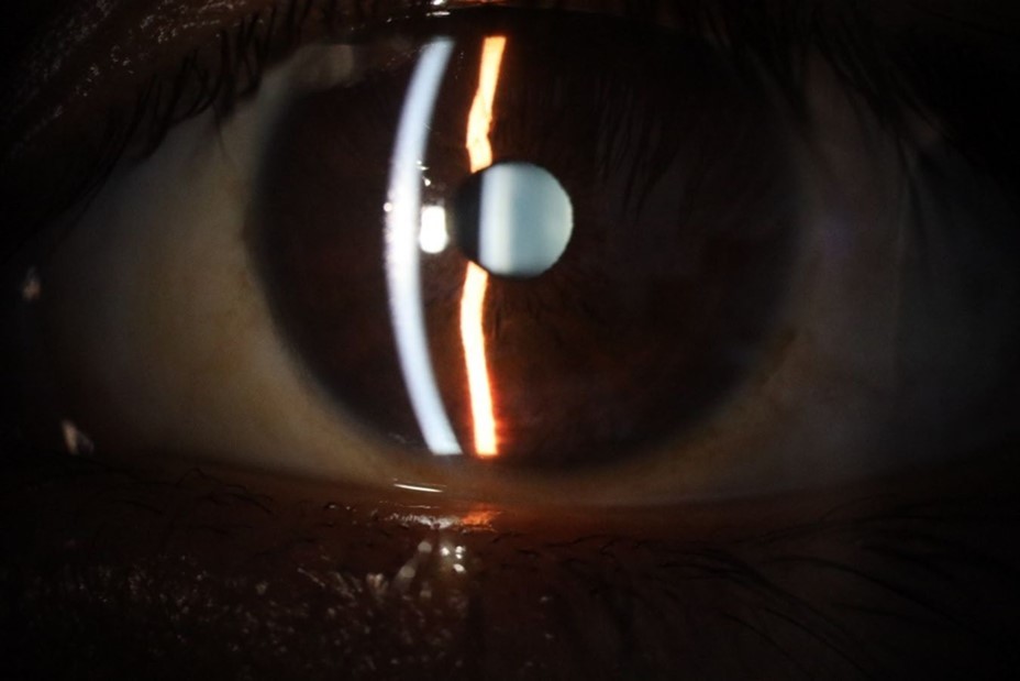
Figure 30.The anatomy of the posterior pole of the left eye is preserved, without showing pathology data.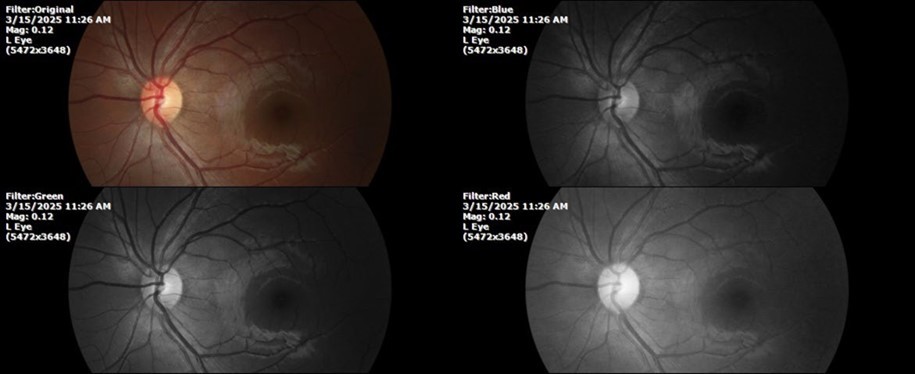
August 26, 2025
It's been fine, there's only a little veil that prevents it from seeing well completely, although it's very transparent (Figure 31, Figure 32, Figure 33, Figure 34, Figure 35, Figure 36).
94 %
62 x ́
++/++
Figure 31.The photograph of the right eye shows an almost normal mirror reflection, as well as a better pupillary dilation.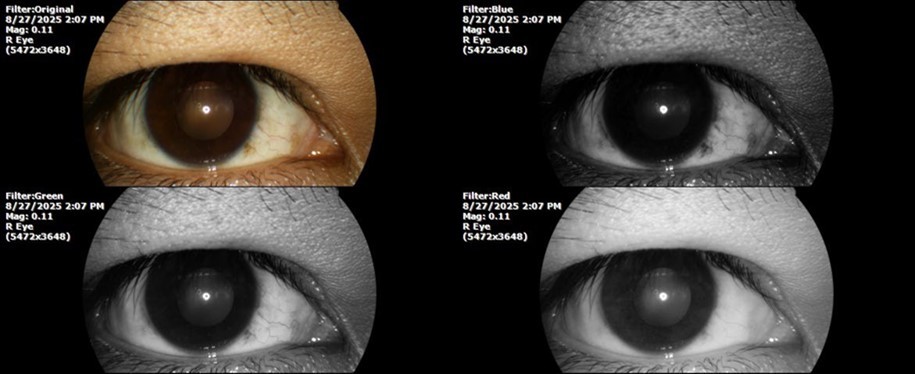
Figure 32.The macro photograph of the right eye shows us a cornea, anterior camera, and crystalline lens in very good condition.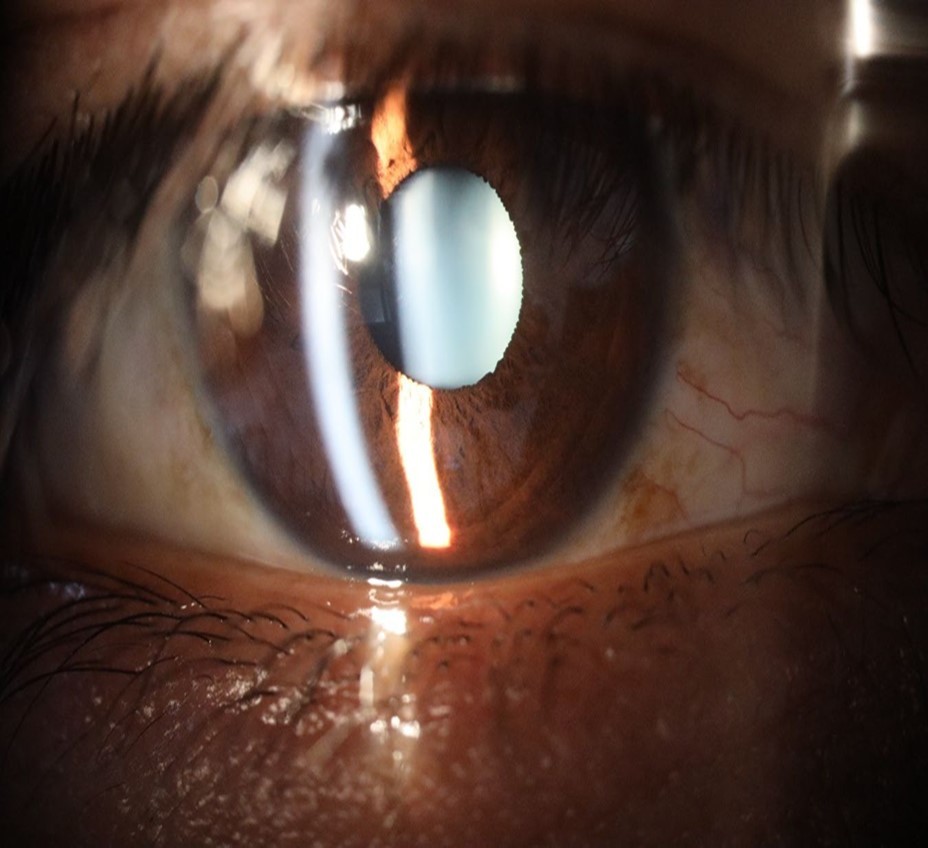
Figure 33.The 3 previous photographs show that whitish mass has almost completely disappeared, and to date only a remnant remains on the left side of the photograph. The chorioretinal scar that is now observed corresponds to the area affected by the blunt trauma, which presumably caused rupture of Bruch's membrane. Fortunately, the macular region has recovered almost in its entirety, so the impairment in central vision was minimal.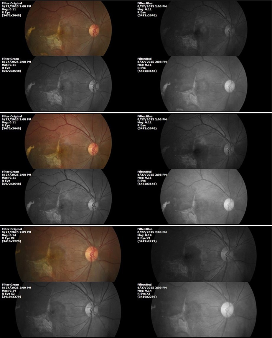
Figure 34.The mirror reflection of the healthy eye (L. E.) continues within normal limits.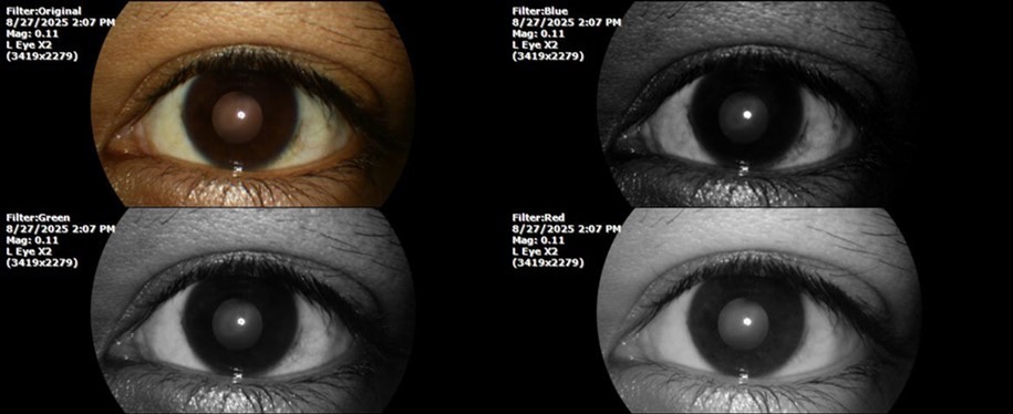
Figure 35.The anatomy of the anterior segment of the left eye is shown without alterations.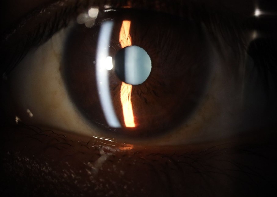
Figure 36.The retina, optic nerve, choroid, and macula of the left eye do not show any evidence of sympathetic ophthalmia.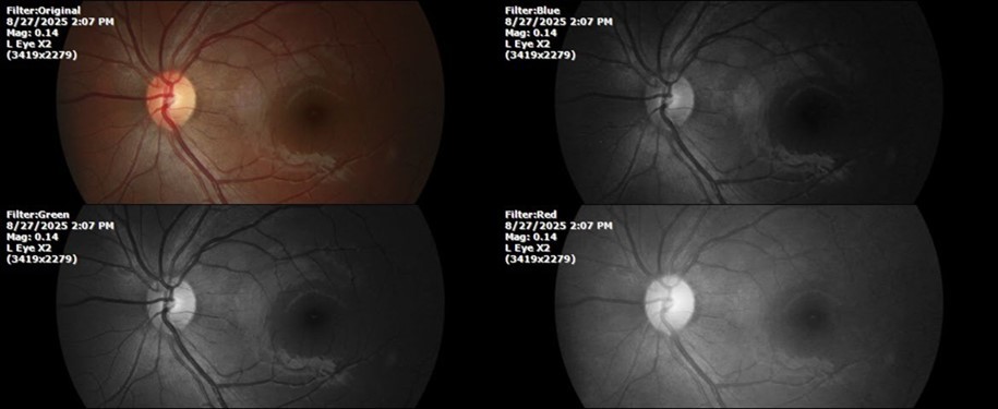
Comment
Despite being a patient affected in the macular region by the blunt trauma he suffered at work, the recovery was very satisfactory, which is not easy to achieve with established orthodox treatments, including surgery and powerful anti- inflammatories.
Conclusion
Commotio retinae with foveal involvement, also known as Berlin´s Edema 7, It is a common cause of monocular vision loss, especially in young male adults. The prognosis is uncertain depending on variables such as location of the affected area, severity of the damage that occurred, extent of the bleeding, vision at the beginning of treatment, etc. There is no single or optimal treatment as it depends on the conditions at the beginning of treatment.
The purpose of publishing this case is to demonstrate the benefits of a new therapeutic approach based on the unsuspected ability of human cells to produce their own oxygen 8, which breaks into a thousand pieces the old dogma, widely spread, that our body took the oxygen it requires for its metabolism from the air that surrounds it.
Acknowledgements
This work was supported by an unrestricted grant from Human Photosynthesis® Research Center, in Aguascalientes 2000, México.
References
- 1.American Academy of Ophthalmology. Basic and Clinical Science Course, section 12: Retina and Vitreous. Leo: he Eye M.D. Association: Posterior Segment Manifestations of Trauma;. 2013-2014.
- 2.Blanch R J, Good P A, Shah P, Bishop J R, Logan A et al. (2013) . Visual outcomes after blunt ocular trauma. Ophthalmology.120(8): 1588–G1. doi: 10.1016/j.ophtha.2013.01.00G. PMID: 23618228.
- 3.Congdon N G, Friedman D S, Lietman T. (2003) Important causes of visual impairment in the world today. Review. PMID: 1455GG61 , JAMA 0(15), 2057-60.
- 5.Souza-Santos F, Lavinsky D, Moraes N S, Castro A R, Cardillo J A et al. (2012) Spectral-domain optical coherence tomography in patients with commotio retinae. , Retina 32(4), 711-8.
- 6.Oladiwura D, Lim L T, Ah-Kee E Y, Scott J A. (2014) Macular optical coherence tomography findings following blunt ocular trauma. Clin Ophthalmol. 8:G8G–G2. doi: 10.2147/OPTH.S64082. PMID: 248GG7G5 .